understanding the importance of nerve protection
When surgeons embrace nerve protection as a part of their algorithm in the OR, they give their patients a better chance of functional recovery and improved quality of life.
why nerves need protection after an injury
Because nerve damage can start from the inside out, an injury isn’t always obvious.
When a traumatic injury occurs, nerves near the site of impact may remain intact and appear outwardly normal. But damage often happens within the nerve or to the tissue surrounding it. Stretching and compressive forces of an injury can cause nerves to experience endoneurial and perineurial damage. In addition, a nerve that appears normal after trauma may become injured over time due to compression from the buildup of scar tissue in the environment surrounding the nerve.
- A deep dive into nerve protection:
- how inflammation occurs
- why protect
- promote healing
- injury mechanism matters
- when to protect
- solutions
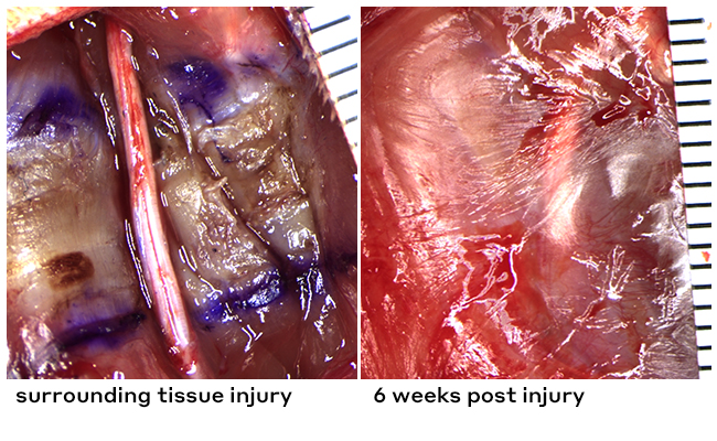
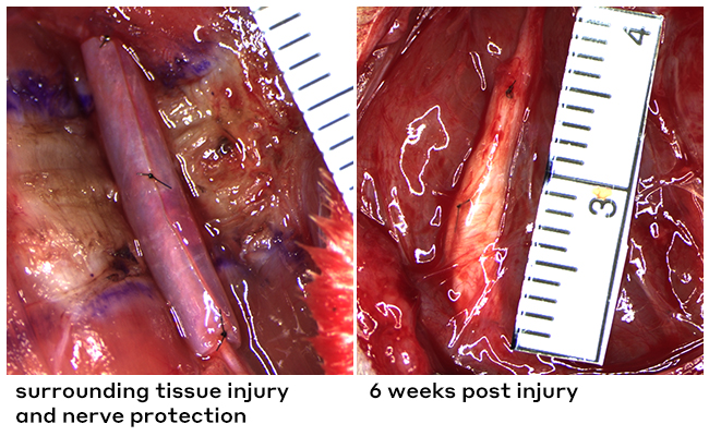
In a thermally injured tissue bed, Axoguard HA+ Nerve Protector™ acts as a physical barrier and protects the nerve from fibrotic tissue
These injuries are sometimes referred to as non-transected nerve injuries (NTNIs). While the nerve may not have been transected, they can lead to the same pain and loss of function if left untreated. One study demonstrated that 69% of hand trauma patients whose nerves were noted to be normal or non-injured at the time of surgery presented signs of impaired nerve function at follow-up.1
Muscle injury: no wrap
Images of rat sciatic nerve, Axogen data on file.
Muscle injury: with wrap
Images of rat sciatic nerve, Axogen data on file.
injured nerves in continuity still need intervention
Despite complete nerve continuity preservation, stretching and compression injuries can result in total loss of both motor and sensory nerve functions. This presents a surgical dilemma due to a lack of obvious repair needed—despite an injury being present.1
77% of patients with hand/wrist trauma and subsequent nerve symptoms are not experiencing full nerve recovery.1
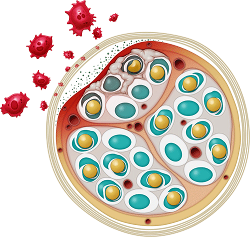
When a nerve is injured, it goes through a healing process in which inflammatory macrophages are recruited to the site of injury to play a critical role in helping to repair the damaged tissue and restore homeostasis.
However, when there is ongoing irritation, such as a nerve rubbing against a plate or tethering due to scar tissue formation, that inflammation can become chronic and lead to additional scarring and pain. These irritations can occur because of damage to the tissue surrounding the nerve, or other factors unique to the patient’s injuries and anatomy.
Types of inflammation in nerve repair

Acute
A part of the normal physiology
Can become chronic with stress or inflammatory triggers in the microenvironment
Chronic
Prolonged inflammation
Can be caused by complex injury or successive acute injuries
May lead to soft-tissue tethering
Nerve protection preserves and protects nerves that may not have been cut or have obvious damage by helping to mitigate soft tissue attachment and block inflammatory macrophage invasion. Introducing a barrier between the non-transected nerve and its surrounding soft tissue creates a safer space for proper nerve regeneration.
In one study, with the absence of direct nerve treatment only 23% of patients experienced full recovery based on qualitative sensory assessments.1
what’s at risk when nerves aren’t protected
- Scarring1,2
- Soft tissue attachment1,2
- Compression3
- Tethering3
- Edema1,2
- Reactive swelling1,2
- Further injury1,2
- Limited mobility1,2
- Decreased sensation1,2
- Incomplete recovery1,2
how nerve protection helps
- Provides a protective barrier to separate the nerve from the surrounding tissues,2,4 minimizing the potential for soft tissue attachments to allow the nerve to glide2
- Reduces the likelihood of traumatized nerve becoming inflamed by serving as a barrier to inflammatory macrophages5
- Reduces the potential for nerve tethering and neurostenalgia3
- Reduces the likelihood of collagen deposition (internal scarring) in the nerve5
When there is trauma to structures adjacent to nerves, this not only hints at the force and mechanism of the injury’s origin but is highly indicative of the likelihood that nerves were involved. When surrounding structures such as ligaments and tendons are torn or transected, it is likely that the adjacent nerve is damaged as well.1
This common correlation highlights the importance of considering the pattern of injury when evaluating for possible nerve involvement.
provide a barrier to inflammatory cells and avoid soft tissue attachment
Adding a barrier between the non-transected nerve and the surrounding soft tissue gives the nerve a better chance at recovery by minimizing the possibility of soft tissue attachments, inflammatory macrophage invasion, and intraneural collagen deposition.
Minimizing soft-tissue attachment is essential because a nerve requires a healthy surrounding tissue bed to allow free nerve gliding.6
the right biomaterial for the right application
Not all barriers are created equal.
build a targeted barrier between nerves and soft-tissue attachment
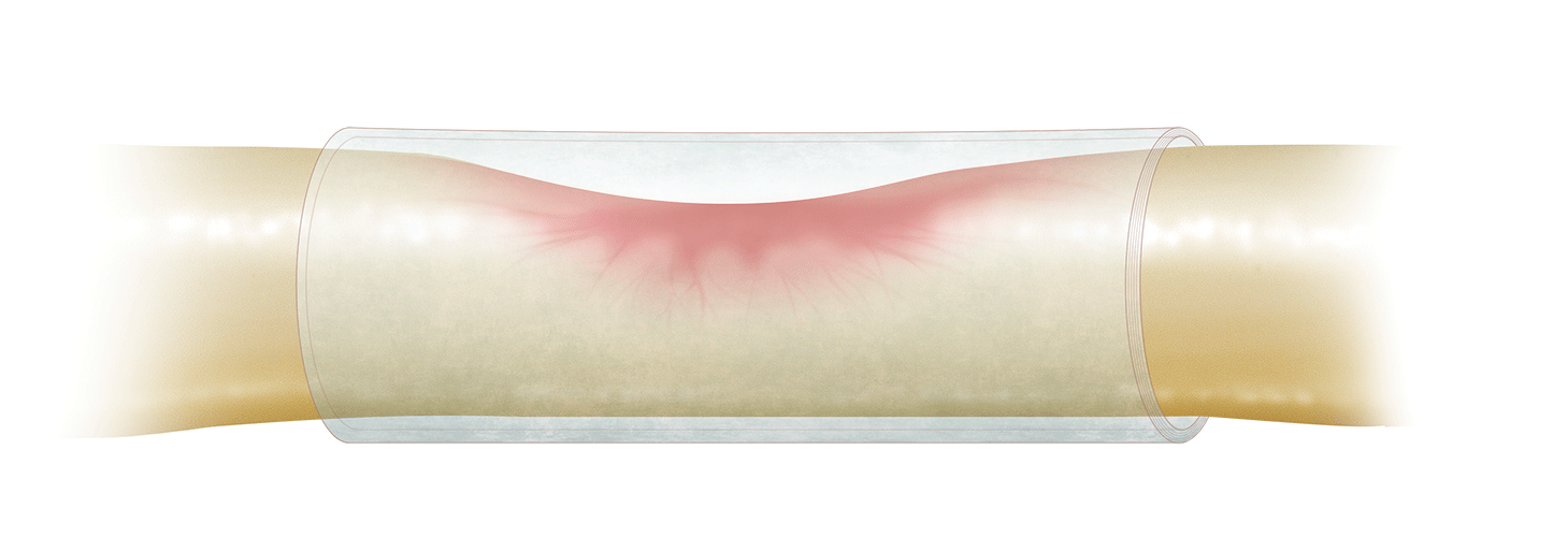
Scar tissue formation in the vicinity of a peripheral nerve can lead to damage, such as compression, ischemia, and impaired glide and tether, which can result in neurostenalgia. Neurostenalgia is a neuropathic pain that results from continuing irritation of an anatomically intact nerve by a noxious agent.6
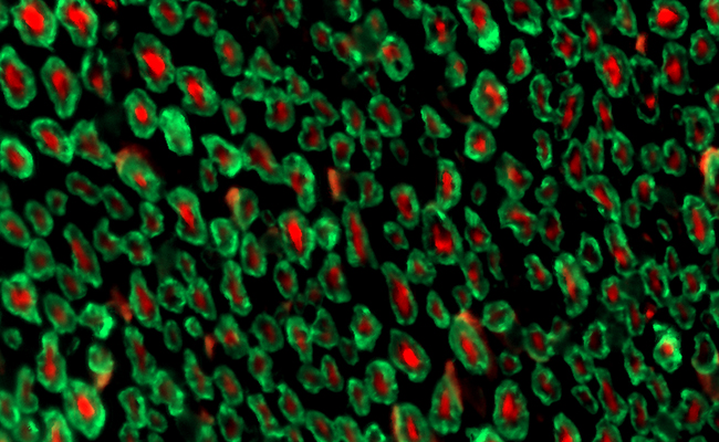
healthy normal nerve fibers
axons: red | myelin: green
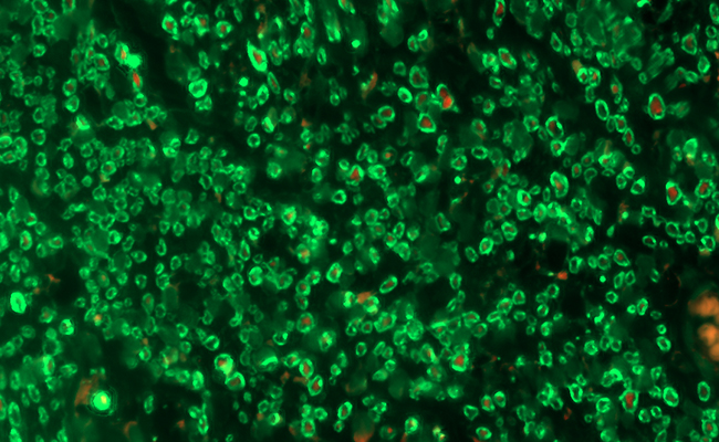
smaller fibers: nerve injury
axons: red | myelin: green
With proper nerve protection, healthy, normal axons can grow more easily. As shown in the left image above, the red staining represents the axons present in a healthy, normal nerve. Conversely, the image on the right represents a nerve that was sitting in an injured muscle bed and wasn’t protected. It shows little to no visible red staining because those axons have been damaged and aren’t there.6
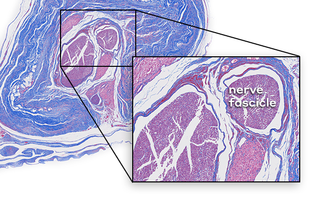
When nerves are properly protected, less collagen buildup occurs. Collagen presence is often indicative of scarring.5
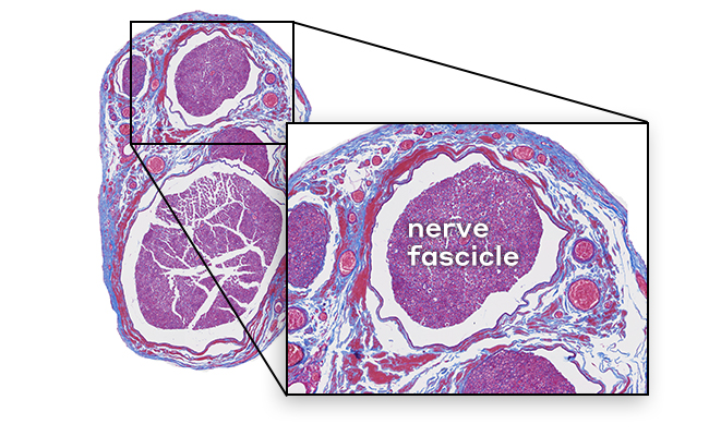
healthy epineurium and fascicles
cells: red | collagen: blue

excess intraneural collagen (unprotected nerve in damaged tissue bed)
cells: red | collagen: blue
minimize collagen deposition (protected nerve in a damaged tissue bed)
cells: red | collagen: blue
Understanding where and why non-transected nerve injuries present themselves, especially in acute hand and wrist trauma, can help in early intervention. And early intervention can help maximize functional recovery.
In the study “Nerves in Continuity Following Hand Trauma: A Descriptive Report,” both injury cause and type were associated with nerve involvement or damage. For example, tendon and ligament injuries were associated with an increased chance of nerve involvement (81.8% and 83.3%, respectively).1
- injury type
- injury mechanism
percentage of patients with abnormal nerve symptoms by injury type1
Tendon and ligament injuries were associated with an increased chance of nerve involvement (81.8% and 83.3%, respectively).1
percentage of patients with abnormal nerve symptoms by injury mechanism1
Blast injuries (such as from a firearm), crush injuries and compression injuries (such as from machinery) are strongly associated with nerve involvement.1
The study featured a retrospective cohort of 20 patients with hand or wrist trauma that presented at the initial surgery with intact nerves. Of these patients, 75% had signs of impaired nerve function either before surgery or at their first post-surgical follow-up. Only 23% experienced full sensory recovery and 57% had full recovery from pain without direct nerve treatment.1
nerves in continuity following hand trauma: a descriptive report–study design
The study featured a retrospective cohort of 20 patients with hand or wrist trauma that presented at the initial surgery with intact nerves. Of these patients, 75% had signs of impaired nerve function either before surgery or at their first post-surgical follow-up. Only 23% experienced full sensory recovery and 57% had full recovery from pain without direct nerve treatment.1
Ensuring patients have the best possible chance for optimal recovery is always the goal. Because of this, protection should be top of mind whenever performing cases where a nerve is involved.
When it comes to nerves, don’t wait to see what transpires after acute surgical healing. Proactive protection sets patients up for long-term functional recovery.
nerve protection is beneficial for:
hardware-based procedures
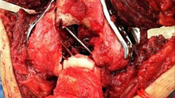
Study results have clearly demonstrated the presence of marked inflammation and tissue reaction in the soft tissue covering stainless steel and titanium plates. A majority of the tissues around metal implants had discoloration and metal fragments within the tissues and macrophages. This is shown to cause immune-inflammatory reactions.7 When near a plate, nerves can rub against the plate surface and incur mechanical irritation directly.
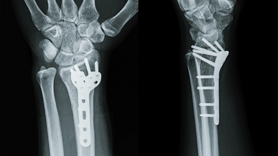
fractures
In one study, 67% of hand fractures presented alongside signs of associated nerve injuries.1 For humerus fractures, fracture location is an important indicator of the probability of a nerve issue. Middle and middle-distal humerus fractures have been most commonly associated with nerve injury.8
The radial nerve is most likely to be damaged in humerus fractures. Especially those that have a lateral displacement of the distal fracture segment, as the nerve is tethered to the bone and cannot withstand the forces applied to it as a result of the displacement.9
radial nerve palsy likelihood based on fracture type and location8
- Transverse: 21.2%
- Spiral: 19.8%
- Oblique: 8.4%
- Comminuted fractures: 6.8%
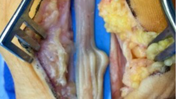
staged and recurrent procedures
Postoperative scarring is the most significant cause of recurrent carpal tunnel syndrome (CTS). In addition, revision surgery is often associated with increased technical difficulty due to significant scarring of the nerve and its surrounding soft tissue.10
Although some reports provide good results with neurolysis alone, many favor providing vascularized tissue or nerve wraps after rerelease to prevent the scarring process. Data suggests nerve protection could provide significant reduction in symptom severity and function severity scores after 1 to 2 years.10
other procedure types
Nerve protection can provide an ideal solution across surgical categories including decompressions, other trauma, head and neck, and iatrogenic injuries.
The following procedures may benefit from using nerve protection, especially Axoguard HA+ Nerve Protector™:
- Spinal accessory nerve after neck dissection
- Radial nerve laying on metal plating for humeral shaft and olecranon fractures
- Ulnar nerve during a revision cubital tunnel release or transposition
- Median nerve through the carpal tunnel in revisions
- Traumatic injuries of the sciatic nerve
- Common peroneal nerve after tibial plateau fractures
- Tibial nerve through tarsal tunnel in revisions
Protecting damaged nerves during the healing process is important for proper healing and consistent patient outcomes. Learn how our solutions help protect nerves from soft tissue attachments and mechanical irritation.
![]()
Remodeling extracellular matrix for long-term protection with a short-term gel coating for enhanced nerve gliding and minimization of soft tissue attachments.
![]()
Extracellular matrix that remodels to protect injured nerves and reinforce nerve reconstructions.
connect with a nerve rep
There’s only a short form between you and our nerve product team who can help you get more information about our nerve repair solutions.
educational events
Axogen offers a variety of events and opportunities for you to interact with experts and stay up to date on the most recent happenings in peripheral nerve surgery.
Sign up for our newsletter to get the latest research, news and in-depth insights about nerve repair.
references
- Pereira DE, et al. Nerves in Continuity Following Hand Trauma: A Descriptive Report. Hand (N Y). 2023;18(1_suppl):126S-132S. doi:10.1177/15589447211064362
- Kokkalis ZT, et al. Assessment of processed porcine extracellular matrix as a protective barrier in a rabbit nerve wrap model. J Reconstr Microsurg. Jan 2011;27(1):19-28. doi:10.1055/s-0030-1267379
- Imran R, et al. Clinical outcomes following neurolysis and porcine collagen extracellular matrix wrapping of scarred nerves in revision carpal tunnel decompression. J Plast Reconstr Aesthet Surg. 2022;75(8):2802-2808. doi:10.1016/j.bjps.2022.04.010
- Thomson SE, et al. Bioengineered nerve conduits and wraps for peripheral nerve repair of the upper limb. Cochrane Database Syst Rev. 2017;2017(3):CD012574. doi:10.1002/14651858.CD012574.pub2
- Axogen data on file.
- Jordaan PW, et al. Management of the scarred nerve using porcine submucosa extracellular matrix nerve wraps. J Musculoskelet Surg Res. 2019;3:128-133.
- Voggenreiter G, et al. Immuno-inflammatory tissue reaction to stainless-steel and titanium plates used for internal fixation of long bones. Biomaterials. 2003;24(2):247-254. doi:10.1016/S0142-9612(02)00312-5
- Shao YC, et al. Radial nerve palsy associated with fractures of the shaft of the humerus: a systematic review. J Bone Joint Surg Br. 2005;87(12):1647-1652. doi:10.1302/0301-620X.87B12.16132
- Bounds EJ, Frane N, Jajou L, Kok SJ. Humeral Shaft Fractures. In: StatPearls. Treasure Island (FL): StatPearls Publishing; March 1, 2023.
- Saffari TM, et al. Compression Neuropathies. Hand Clinics. 2023;39(3):389-401. doi:https://doi.org/10.1016/j.hcl.2023.02.009